Header

Search

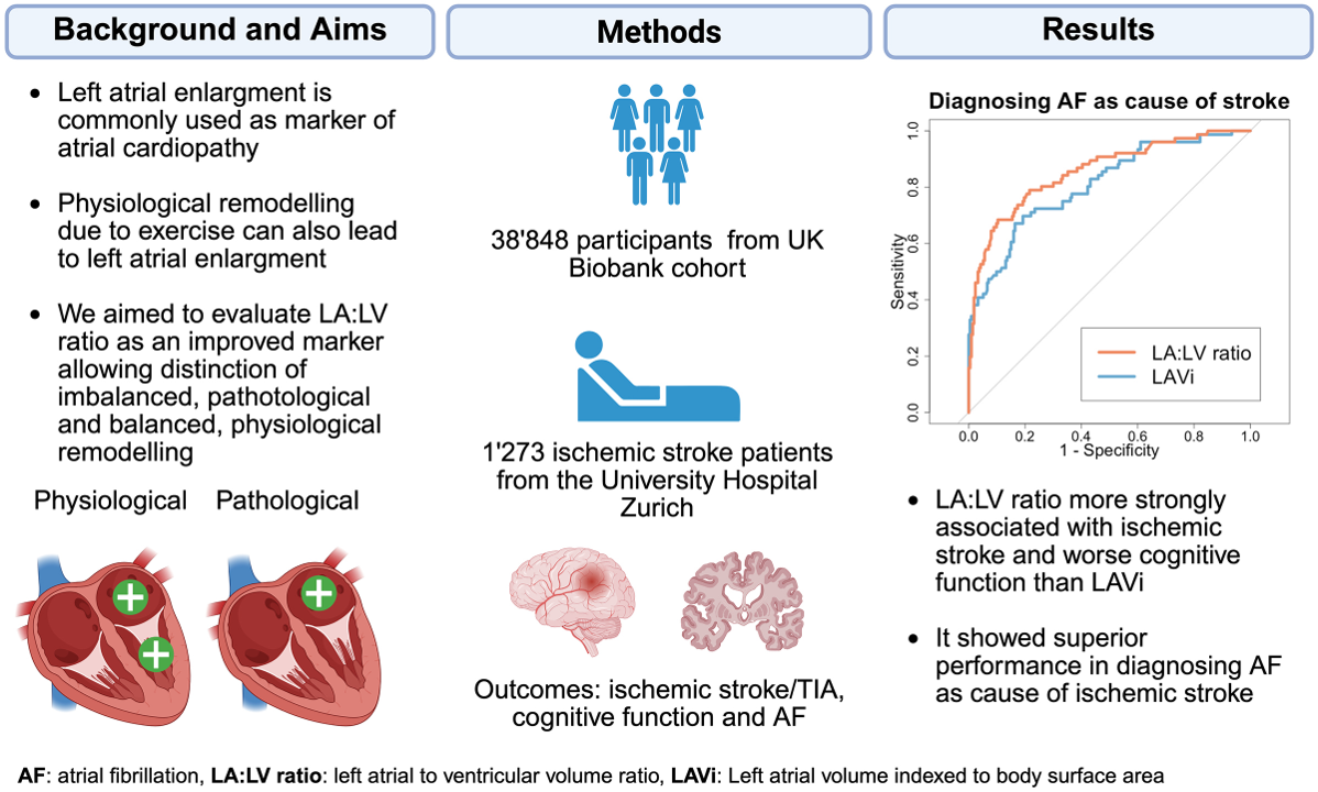
‎Improving diagnosis of cardioembolic stroke with machine learning
https://pubmed.ncbi.nlm.nih.gov/42312392/

By analyzing brain magnetic resonance imaging, cardiac imaging and electrocardiogram using machine learning techniques, we aim to improve the detection of patients with a cardioembolic cause of stroke for targeted prophylaxis of recurrence using anticoagulation.

Ratio of Left Atrial and Ventricular Volume as New Marker of Atrial Cardiopathy and Stroke Risk - PubMed

Deep learning enables diagnosis of atrial cardiomyopathy from routine 12-lead electrocardiogram | medRxiv

Furthermore,  we work on deep learning models that enable extraction of features predicting imaging readouts of atrial cardiomyopathie from routine ECG (even reduced lead holter ECG).  This may help early identification of those at risk of stroke, heart failure and atrial fibrillation.


People involved: 

Julian Deseö, Lisa Herzog, Hakim Baazaoui, Prof. Bjoern Menze, Ezequiel de la Rosa (DQBM)
Team

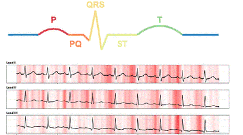
Importance of different ECG segments calculated using vanilla gradients for prediction of indexed left atrial maximum volume (A), indexed left atrial minimum volume (B), left atrial ejection fraction (C) and left atrial to ventricular volume ratio (D) evaluated on 1’077 5 second non-AF ECG samples from 653 patients from the USZ stroke cohort over all 12 leads. Importance was calculated by averaging absolute saliency values over all 12 leads per segment and then dividing by average salience of the entire 5 second ECG sample. (E) Schematic representaion of the ECG segments evaluated in (A)-(D). (F) Example saliency map for leads I-III. Darker red indicates high absolute saliency. LA max: indexed left atrial maximum volume, LA min: indexed left atrial minimum volume, LAEF: left atrial ejection fraction, LALV: left atrial to ventricular volume ratio.